Hot: UHF Small Animal Doppler Lab Animal Ultrasound Portable Pet Doppler Handheld Vet Ultrasound

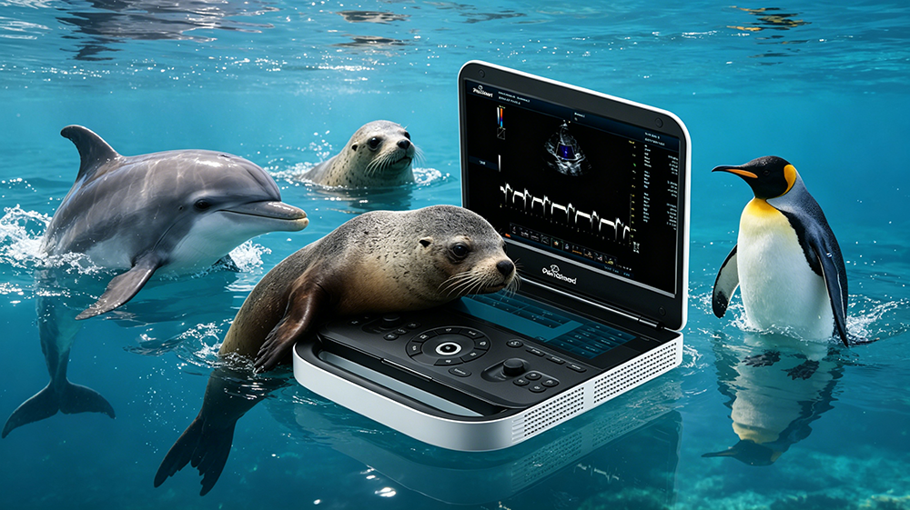
Periodmed's animal ultrasound system provides professional medical care for marine life such as dolphins, sea lions, and penguins. Aquariums, marine conservation institutions face unique challenges in managing the health of marine animals like dolphins, sea lions, walruses, and penguins during their daily operations. These species vary widely in size, live in special environments, and are highly sensitive to stress. Traditional examination methods often have limitations such as being highly invasive and difficult to operate. Periodmed's animal-specific ultrasound system, with its non-invasive, real-time, and precise imaging advantages, provides a professional solution for examining abdominal organs and monitoring pregnancy in marine animals.
I. Core Application Scenarios
1. Routine Health Monitoring: For large marine mammals such as dolphins and sea lions, the equipment supports rapid abdominal organ screening in medical pools or temporary holding areas, assessing the structural and functional status of internal organs such as the liver, kidneys, and gastrointestinal tract, and establishing individual health baseline data.
2. Pregnancy Monitoring and Breeding Management: For pregnant dolphins, sea lions, walruses, and other species, ultrasound can be used to track fetal development in real time, monitoring key indicators such as fetal heart rate, gestational sac size, and placental blood flow, optimizing perinatal care plans, and improving calf survival rates.
3. Examination of Small and Medium-Sized Marine Birds such as Penguins: Given the small size and high metabolic rate of penguins, the high-frequency probe can clearly visualize the fine structures of their abdominal organs, assisting in the diagnosis of common problems such as digestive system diseases and reproductive system abnormalities.
4. Injury and Illness Diagnosis and Rehabilitation Tracking: For postoperative animals or individuals with injuries or illnesses, regular ultrasound follow-ups can dynamically observe the progress of tissue healing, fluid absorption, and blood flow reconstruction, providing objective evidence for adjusting rehabilitation plans.

II. Key Examination Items
Abdominal Organ System Assessment
Liver: Observe the homogeneity of parenchymal echoes, vascular course, and space-occupying lesions.
Kidneys: Assess the cortical-medullary boundary, renal pelvis structure, and stones/cysts.
Gastrointestinal Tract: Monitor gastric contents, intestinal wall thickness, and peristaltic function.
Spleen/Pancreas: Screen for enlargement, inflammation, or signs of tumors.
Reproductive and Obstetric Specialized Examinations
Pregnancy Diagnosis: Early gestational sac localization and confirmation of embryonic viability.
Fetal Development Monitoring: Quantify growth parameters such as biparietal diameter, body length, and heart rate.
Maternal Reproductive System: Screen for pathological conditions such as ovarian cysts and endometritis.
Placement Blood Flow Assessment: Utilize ultra-microscopic blood flow imaging technology to observe the distribution of microvessels at the 10-100 micrometer level and assess placental perfusion status.
Cardiovascular Function Auxiliary Assessment
For mammals such as dolphins and sea lions, echocardiography can be performed simultaneously to assess cardiac chamber structure, valve opening and closing, and hemodynamics, and to screen for heart disease risks.
III. Technological Advantages and Adaptability to Marine Scenarios
1. Clear Imaging of Deep Tissues: Large animals such as dolphins and walruses have thick abdominal walls. The device, equipped with a 2MHz low-frequency probe and a high-sensitivity signal receiving system, can effectively penetrate deep tissues, accurately capture weak signals, and ensure stable and reliable imaging quality for deep organs such as the liver and kidneys.
2. Rapid Physiological Motion Capture Without Motion Artifacts: Marine mammals have fast heart rates. The device uses an ultra-high frame rate imaging algorithm (>500fps) to track rapid physiological processes such as heartbeats, fetal heart activity, and blood perfusion in real time, effectively avoiding motion artifacts and accurately reproducing dynamic images.
3. High-Resolution Representation of Fine Structures: For small and medium-sized species such as penguins, ultra-high resolution imaging at the 50-100 micrometer level can clearly present the fine structures of their organs, aiding in early lesion identification and accurate diagnosis.
4. Portable Design Adaptable to Diverse Environments: The device features a lightweight and portable body, supports battery power, and can be flexibly deployed near medical pools, in isolation areas, or during transport, meeting the diverse examination needs of aquariums. IV. Welfare Value for Marine Life
1. Non-invasive, Low-stress Examination: Ultrasound examinations require no surgery or puncture and can be completed quickly with professional restraint, significantly reducing animal stress and anesthesia risks, meeting modern animal welfare standards.
2. Early Disease Warning: Regular ultrasound screening can detect potential problems such as organ structural abnormalities and microcirculatory disorders before clinical symptoms appear, achieving "early detection, early intervention," improving treatment outcomes and survival rates.
3. Scientific Breeding Management: Quantitative assessment of pregnancy status and fetal development optimizes mating plans, prenatal nutrition, and parturition plans, improving population breeding success rates and genetic diversity management.
4. Traceable Health Records: Supports DICOM format data export and standardized report generation, facilitating the establishment of individual full-lifecycle health records, providing objective data support for cross-year trend analysis, scientific research collaboration, and conservation decisions.
V. Implementation Recommendations
Before formal implementation, aquariums are advised to complete the following preparatory work: ① Clearly define the target species list and core examination areas; ② Have veterinarians with experience in aquatic animal ultrasound perform the procedure, using professional coupling media and waterproofing measures; ③ Establish standardized examination procedures and data archiving systems; ④ Conduct regular operational training and image quality assessments.
The Periodmed animal ultrasound system, with its professional imaging technology and flexible scene adaptability, provides reliable technical support for marine animal health management, contributing to the scientific development of marine conservation.
Technical Support: Providing marine animal ultrasound examination operation training and customized measurement solutions.
【Editor】admin
 Address
13th Floor, Building C1,Mingyang Square,Software Park Economic development DistrictXuzhou
 Email
Email
info@periodmed.com
 Tel/Whatsapp
+86 15005204265


