

动物彩超诊断仪
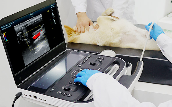
The Revo 9 VET is a portable color Doppler ultrasound diagnostic system designed specifically for clinical diagnosis and treatment of research animals. It supports ultra-high frequency imaging up to 38MHz, integrates a high-element channel architecture, a GPU soft beam computing platform and a dual-screen interactive design, and integrates multi-modal imaging modes and AI intelligent algorithms. It aims to provide veterinary clinics with efficient, accurate and easy-to-use image diagnostic support.
Tel/Whatsapp:+86 15005204265
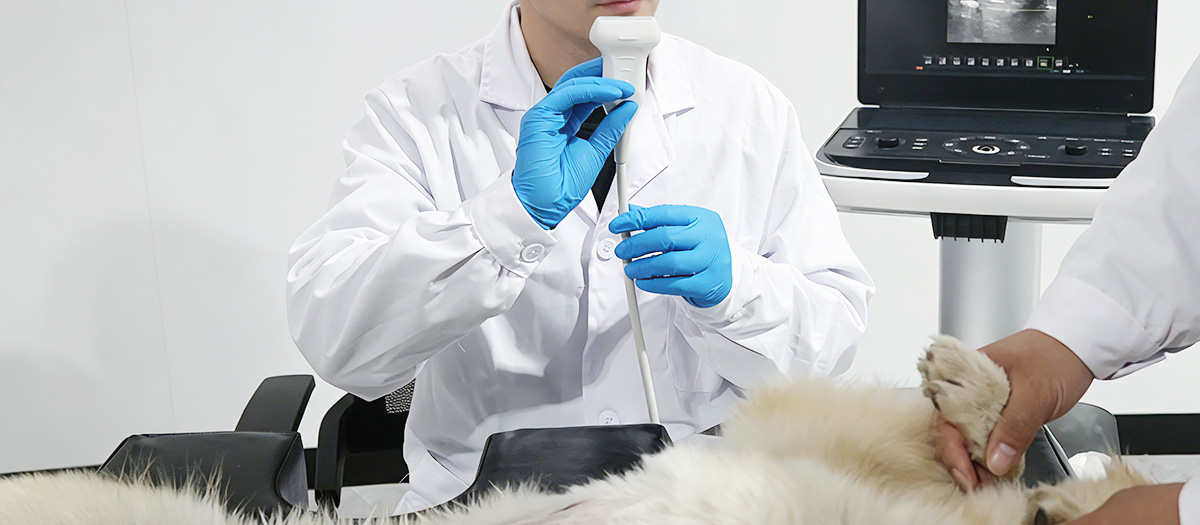
The Revo 9 heart ultrasound system is conceived to optimize cardiac examination workflows, transforming daily clinical routines into a highly productive platform. By streamlining operations and reducing scanning time, this advanced system empowers clinicians to focus more on patient care without compromising on diagnostic efficiency.
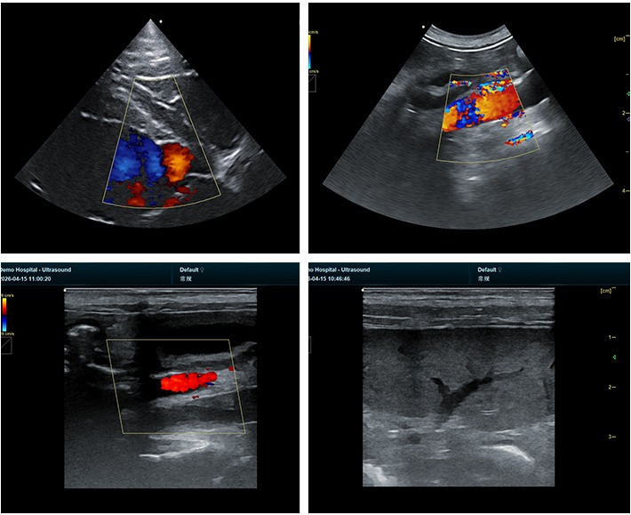
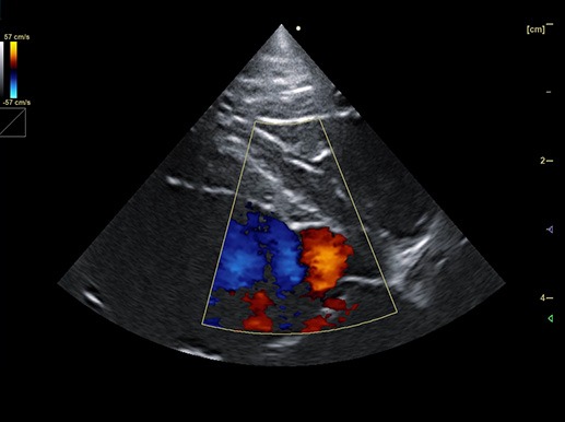
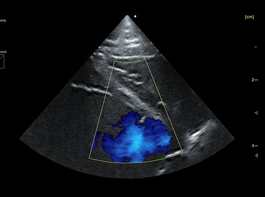
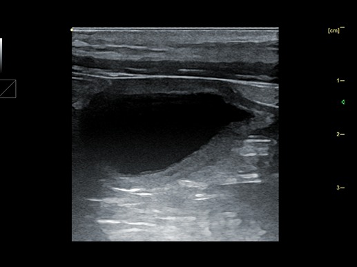
Utilizing advanced sound wave technology, our heart ultrasound device captures real-time, high-definition images of the patient's heart to evaluate its structure, function, and blood flow. This non-invasive procedure provides crucial insights, enabling physicians to precisely assess and manage complex cardiac conditions, including heart failure, valvular regurgitation, and heart murmurs.

Dual-screen independent operation: A 15.6-inch main display + a 12.6-inch touchscreen allow for focused scanning on one screen and parameter adjustment on the other, without interference, significantly improving scanning efficiency; Dual probe interfaces: No need to plug or unplug, freely switchable, highly efficient and convenient, with an optional triple probe interface for expanded applications.
Built-in AI intelligent algorithm: reduces the difficulty of operation and shortens the diagnosis and treatment time; intelligent image optimization: automatically identifies standard cross-sections and optimizes image parameters, significantly reducing the reliance on operator experience.
Advanced Imaging Suites and Diagnostic Modes
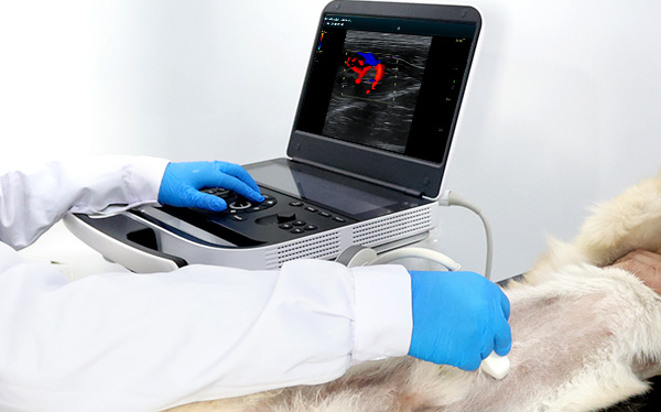
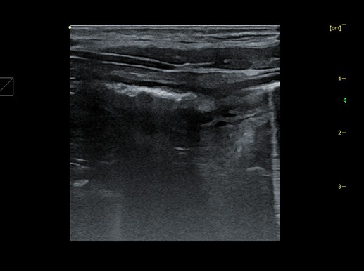
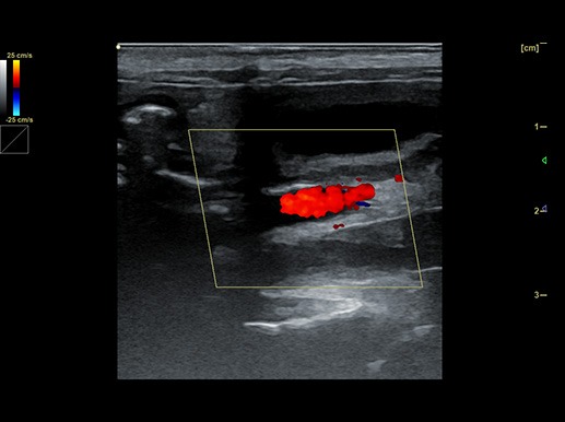
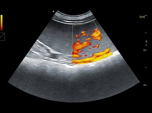
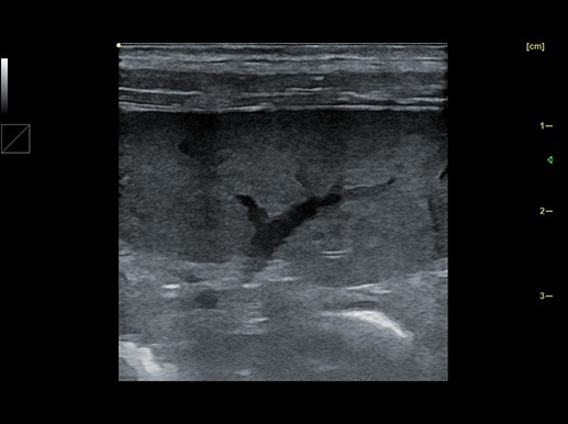
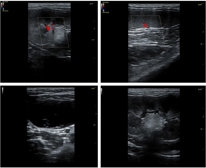
The system is equipped with a comprehensive suite of advanced imaging modes designed to elevate diagnostic confidence across diverse clinical scenarios. Featuring automatic envelope imaging, enhanced needle puncture visualization, and strain elastography, it provides clinicians with precise anatomical and tissue-stiffness insights. Furthermore, cutting-edge modalities such as wide-view imaging, trapezoidal imaging, and high-sensitivity micro-blood flow imaging—alongside state-of-the-art real-time 3D and 4D volume rendering—ensure exceptional detail resolution from superficial structures to complex deep tissues.
Enhanced Visualization and Tissue Contrast
To further optimize image interpretation, the platform offers more than 28 customizable pseudo-color display modes. This extensive color-mapping versatility significantly enhances subtle tissue contrast, allowing sonographers to easily differentiate subtle pathological variations, reduce eye fatigue during extended exams, and achieve unprecedented diagnostic accuracy.
 Address
13th Floor, Building C1,Mingyang Square,Software Park Economic development DistrictXuzhou
 Email
Email
info@periodmed.com
 Tel/Whatsapp
+86 15005204265


