Hot: UHF Small Animal Doppler Lab Animal Ultrasound Portable Pet Doppler Handheld Vet Ultrasound

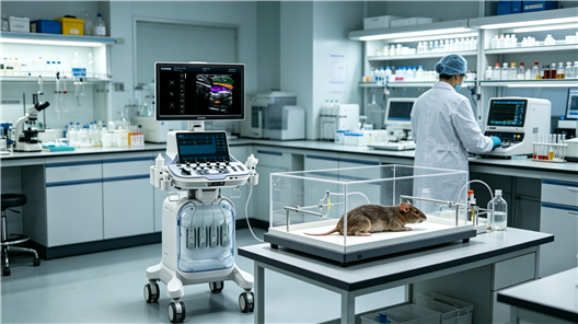
For research institutions and university laboratories engaged in life sciences, translational medicine, and animal husbandry and veterinary medicine, a high-performance animal-specific color Doppler ultrasound machine is a key piece of equipment for non-invasive, in vivo microscopic imaging. This document will provide an in-depth analysis of how small animal ultrasound imaging systems can serve cutting-edge research, focusing on their specialized solutions for mouse cardiac function analysis, liver elastography, hemodialysis, and preclinical studies in non-human primates such as monkeys, offering authoritative references for your equipment selection and research design.
I. Microscopic Imaging and Quantitative Studies in Rodent Models
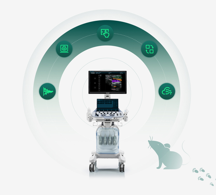
The Promax Animal Color Doppler Ultrasound System is equipped with ultra-high frequency linear and convex array probes (center frequency up to 30MHz), optimized for imaging small organs in mice, rats, and other animals, providing excellent detail resolution.
1. Mouse Heart Structure and Functional Dynamics Studies
• Application Scenarios: Myocardial infarction (MI) models, heart failure models, myocardial hypertrophy models, drug efficacy evaluation.
• Technical Achievements:
◦ High-resolution B/M-mode ultrasound: Clearly observes the thickness of the left ventricular anterior/posterior wall, interventricular septum, and real-time cardiac motion.
◦ Pulsed Doppler (PW) and continuous Doppler (CW): Accurately measures blood flow velocity at the aortic and mitral valve orifices, calculating core cardiac function parameters such as stroke volume and cardiac output.
◦ Tissue Doppler Imaging (TDI): Quantitatively assesses local myocardial motion velocity for earlier detection of cardiac dysfunction.
2. Non-invasive Imaging Assessment of Liver Disease Models
• Application Scenarios: Liver fibrosis/cirrhosis models, non-alcoholic fatty liver disease models, hepatocellular carcinoma xenograft models.
• Technical Implementation:
◦ B-mode ultrasound: Real-time observation of liver morphology, capsule smoothness, parenchymal echo homogeneity, and space-occupying lesions.
◦ Elastography: One of the core functions of this equipment. Quantitative measurement of liver tissue stiffness enables non-invasive, dynamic monitoring of liver fibrosis staging, significantly superior to traditional pathological examination, greatly improving experimental efficiency and animal welfare.
3. Blood Perfusion Research in Oncology and Vascular Biology
• Application Scenarios: Tumor angiogenesis and evaluation of anti-angiogenic drugs, ischemia-reperfusion models (stroke, lower limb ischemia).
• Technical Implementation:
◦ Color Doppler and Energy Doppler: Visually displays the distribution and density of microvessels within tumors or organs.
◦ Contrast-enhanced ultrasound imaging: The second core function of this equipment. Through intravenous injection of targeted or non-targeted microbubble contrast agents, true real-time dynamic visualization of blood perfusion is achieved. Time-intensity curves (TICs) can be generated to quantitatively analyze blood flow velocity, blood volume, and peak intensity, serving as the gold standard for evaluating the efficacy of anti-angiogenic therapy.
II. Preclinical Translational Research in Non-Human Primates (NHP)
• Application Scenarios: Neuroscience (transcranial ultrasound), reproductive and developmental biology, infectious disease models, safety evaluation of macromolecular drugs.
• Technical Advantages: The equipment is compatible with low-to-medium frequency curved probes and is suitable for medium-sized animals such as monkeys and beagles. High frame rate imaging ensures stable image acquisition in awake or mildly sedated animals, meeting the data consistency requirements of long-term longitudinal studies and providing key imaging biomarkers for drug translation from the laboratory to the clinic.
III. Integrated Teaching and Research Applications in Agricultural and Animal Husbandry Veterinary Colleges
• Teaching Value: As an advanced teaching tool for courses such as Veterinary Imaging, Experimental Animal Science, and Comparative Medicine, it enables students to master in vivo ultrasound scanning skills for animals ranging from laboratory mice to economically important animals (pigs and sheep).
• Research Expansion:
◦ Livestock Breeding: Used for dynamic monitoring of ovaries and embryos in breeding livestock, and precise measurement of backfat thickness and eye muscle area in live animals.
◦ Disease Research: Constructing metabolic syndrome and cardiovascular disease models in large animals (such as pigs) and conducting non-invasive follow-up.
Summary: Why choose Promax Animal-Specific Color Doppler Ultrasound System?
Promax is not a simple modification of ordinary medical ultrasound, but an imaging platform deeply customized for scientific research and teaching scenarios. Its integrated high-frequency B-mode ultrasound, Doppler, ultrasound contrast imaging, and shear wave elastography technologies form a complete non-invasive assessment loop from anatomical structure and hemodynamics to tissue biomechanical properties. Whether exploring the mysteries of the heart in genetically modified mice, evaluating the safety of new drugs in monkey models, or training a new generation of veterinary imaging professionals, it is a reliable cornerstone supporting high-level research and producing high-quality data.
【Editor】admin
 Address
13th Floor, Building C1,Mingyang Square,Software Park Economic development DistrictXuzhou
 Email
Email
info@periodmed.com
 Tel/Whatsapp
+86 15005204265


