Hot: UHF Small Animal Doppler Lab Animal Ultrasound Portable Pet Doppler Handheld Vet Ultrasound

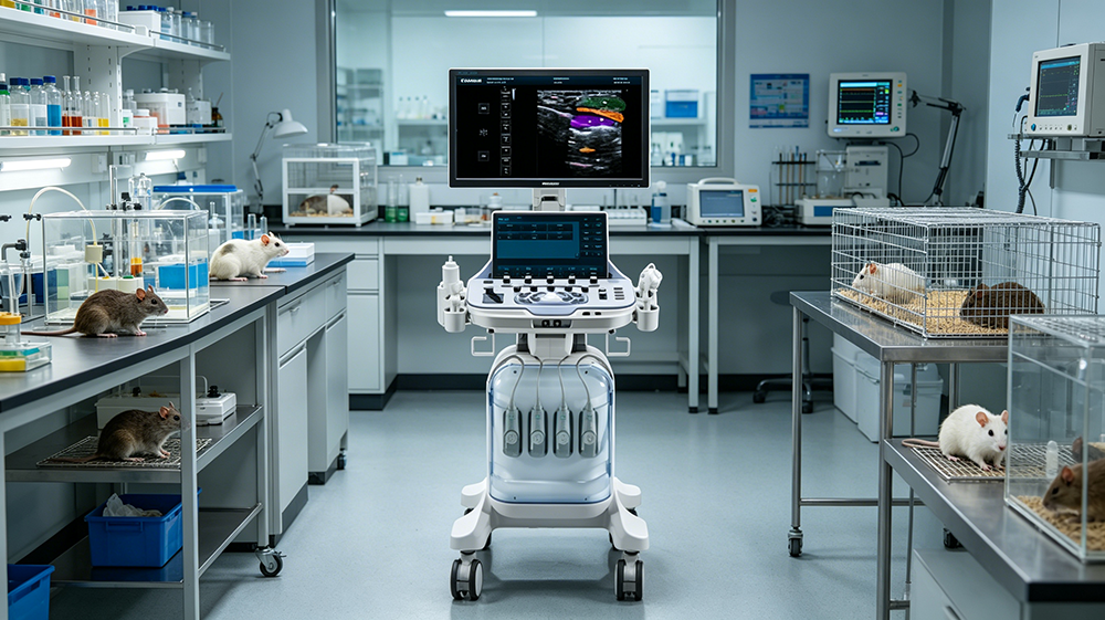
I. Pain Points and Needs of Small Animal Imaging in Scientific Research In biomedical research, mice and rats are the most commonly used experimental models, and dynamic monitoring of their organ functions (such as heart and liver) and disease mechanisms (such as tumors and metabolic diseases) is a core need. Traditional imaging technologies (such as CT and MRI) have limitations such as radiation risks, high costs, and the inability to perform real-time dynamic observation. Animal color Doppler ultrasound, with its advantages of being non-invasive, real-time, repeatable, and low-cost, has become the preferred tool for research institutions. The Revo T2 VET, a cart-type color Doppler ultrasound system specifically designed for animals, features high resolution, a multi-functional probe, and a portable design, precisely matching the research scenarios of mice and rats, providing crucial data support for research on the heart, liver, and tumors.
II. Product Technical Advantages: Core Parameters Supporting Precise Scientific Imaging
The Revo T2 VET's technical parameters directly serve the observation of fine structures in small animals. Its core advantages include:
High-resolution imaging: A 192/64-element/channel probe, combined with 16-beam planar wave technology, enhances image frame rate and clarity, clearly displaying fine tissues such as mouse myocardium (thickness <1mm) and liver microstructures (liver lobules);
Wide dynamic range: A 36-120dB dynamic range adapts to different tissue densities in mice and large animals (e.g., high echogenicity in fatty liver, low echogenicity in normal liver), reducing artifacts and improving lesion recognition rate;
Multi-functional probe compatibility: Equipped with convex array (C5-2R60N), linear array (L12-5L40H), cardiac array (P4-2L15SI), and micro-convex array (Micro-Convex). Probes such as the C5-2R20N cover areas including the heart, abdomen, and pelvis, meeting the needs of multi-organ research; Convenient operation design: trolley-style base (with wheels for movement), 24-inch large screen (-75°~+75° tilt, 90° rotation), 13.3-inch touchscreen (180° rotation), adaptable to laboratory workbench height, reducing animal stress; Advanced imaging modes: elastography (assessing tissue stiffness), contrast imaging (microbubble perfusion analysis), 3D/4D imaging (stereoscopic observation of organ structures), supporting in-depth data mining in scientific research.
III. Core Scientific Application Scenarios: Precision Tools for Mouse and Rat Research
1. Heart Research: Dynamic Monitoring of Myocardial Structure and Cardiac Function
Application Scenarios: Assessment of myocardial thickness, ventricular volume, and ejection fraction (EF) in models of cardiomyopathy, hypertension, and heart failure.
Technical Support: A phased array cardiac probe with high dynamic range can clearly display mouse myocardial layers (e.g., left ventricular wall thickness) and valve motion (e.g., mitral valve opening amplitude); elastography can quantify myocardial stiffness (e.g., degree of fibrosis), aiding in the assessment of disease progression.
Scientific Value: Real-time monitoring of changes in cardiac function reduces the frequency of animal sacrifice, conforming to the "Reduction" 3R principle; high-resolution images support quantitative analysis (e.g., myocardial strain rate), improving the reliability of data in research papers.
2. Liver Research: Evaluation of Fatty Liver, Fibrosis, and Tumor Models
Application Scenarios: High-fat diet-induced fatty liver (increased echogenicity), carbon tetrachloride-induced liver fibrosis (increased tissue stiffness), and tumor growth monitoring in liver cancer transplantation models.
Technical Support: The convex array probe (C5-2R60N) is suitable for imaging the mouse abdomen, offering a wide dynamic range to distinguish the echogenicity differences between normal and fatty liver; elastography can quantify the degree of liver fibrosis (e.g., stiffness value), and contrast imaging (microbubble contrast agent) can observe tumor angiogenesis (e.g., microvessel density).
Research Value: Non-invasive and repetitive monitoring of liver disease progression, replacing traditional histopathology (reducing animal use); contrast imaging can evaluate the efficacy of antitumor drugs (e.g., anti-angiogenic drugs), shortening the experimental cycle.
3. Contrast Imaging: Hemodynamics and Tumor Angiogenesis Research
Application Scenarios: Tumor microvascular perfusion, blood flow restoration in ischemia-reperfusion injury, and efficacy evaluation of antithrombotic drugs.
Technical Support: 16-beam planar wave technology improves the contrast frame rate (up to 50-400Hz), and when used with microbubble contrast agents (such as SonoVue), it allows real-time observation of microvascular perfusion (diameter <100μm) within mouse tumors; Doppler function measures blood flow velocity and resistance index (RI) to analyze vascular stenosis or thrombosis.
Research Value: Visually displays tumor angiogenesis (such as VEGF expression levels), providing visualized data for anti-angiogenic drug development; hemodynamic parameters (such as RI) can predict disease prognosis, enhancing research depth.
4. Tumor Research: Tumor Boundary, Blood Supply, and Metastasis Monitoring
Application Scenarios: Size, boundary, and blood supply analysis of subcutaneous xenografts (such as lung cancer and breast cancer); early detection of metastatic tumors (such as lung metastases).
Technical Support: Linear array probes (L12-5L40H) provide high-resolution display of tumor boundaries (clarity up to 0.1mm); contrast imaging can quantify tumor blood supply (such as perfusion intensity); 3D imaging allows for three-dimensional observation of tumor morphology, assisting in determining the location of metastatic lesions.
Research Value: Non-invasive monitoring of tumor growth (e.g., weekly volume measurement), replacing traditional caliper measurements (reducing errors); blood supply analysis can assess the efficacy of chemotherapy/radiotherapy (e.g., tumor angiogenesis reduction), accelerating drug screening.
IV. Practical Value to Research Institutions: Improved Efficiency, Cost, and Data Quality
1. Increased Operational Efficiency: The cart-style design facilitates movement within the laboratory; the touchscreen and probe rotation function reduce operational steps and shorten single examination time (only 5-10 minutes for mice), increasing experimental throughput.
2. Guaranteed Data Quality: High resolution and wide dynamic range ensure clear images; DICOM format compatibility with laboratory information systems (e.g., LabVIEW, ImageJ) facilitates quantitative analysis (e.g., myocardial thickness, tumor volume); elastography and contrast imaging provide multi-dimensional data, enhancing the competitiveness of published papers.
3. Optimized Cost-Effectiveness: Compared to CT/MRI, animal ultrasound reduces costs by more than 50%; multiple probes allow for multi-purpose use, reducing the number of devices required; non-invasive imaging reduces animal usage (e.g., each mouse can be examined more than 10 times), lowering ethical approval difficulty and experimental costs.
4. Ethical and Compliance Support: Non-invasive, real-time imaging conforms to the 3R principle of "Replacement," reducing animal suffering; data traceability (DICOM storage) meets the requirements of research ethics review.
V. Periodmed's Innovative Engine for Animal Model Research: The Revo T2 VET trolley-type animal ultrasound machine, with its technical precision, ease of operation, and data reliability, has become a powerful tool for mouse and rat research. Its application capabilities covering multiple organs such as the heart, liver, and tumors not only solve the pain points of traditional imaging technologies but also provide research institutions with an efficient, low-cost, and ethically compliant research tool, assisting in the mechanism research and drug development in fields such as cardiovascular diseases, tumors, and metabolic diseases. In the future, with further upgrades in ultrasound technology (such as AI-assisted analysis), the Revo T2 VET will continue to drive the innovative process of small animal model research.
【Editor】admin
 Address
13th Floor, Building C1,Mingyang Square,Software Park Economic development DistrictXuzhou
 Email
Email
info@periodmed.com
 Tel/Whatsapp
+86 15005204265


