Hot: UHF Small Animal Doppler Lab Animal Ultrasound Portable Pet Doppler Handheld Vet Ultrasound

I. Product Overview
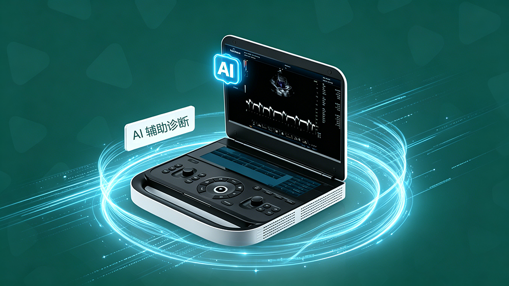
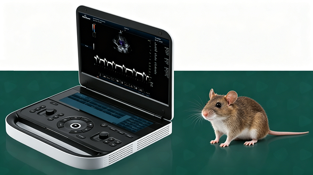
The Revo 9 Vet is a portable color Doppler ultrasound diagnostic system designed specifically for animal clinical diagnosis and treatment. The device integrates a high-element channel architecture, a GPU soft-beam computing platform, and a dual-screen interactive design. It incorporates multimodal imaging modes and AI intelligent algorithms, aiming to provide efficient, accurate, and easy-to-use imaging diagnostic support for veterinary clinics, and is fully adaptable to the examination needs of multiple departments, including cardiology, abdominal surgery, obstetrics, musculoskeletal surgery, ophthalmology, and interventional guidance.
II. Core Imaging Technologies
High-Element Channel and Plane Wave Technology: Equipped with a 192-element/64-channel hardware architecture, combined with rapid sound velocity calibration technology and 16-beam plane wave technology, it provides dynamically accurate calculation parameters, effectively improving beam focusing accuracy and image spatial resolution.
GPU Architecture Soft Beamformer: Relying on underlying high computing power, it provides the foundation for the implementation of new imaging algorithms, fundamentally optimizing ultrasound image quality and ensuring detail reproduction and noise suppression in complex anatomical structures.
Wide Dynamic Range and Depth Coverage: Supports a dynamic range adjustment of 36~120dB, a pulse repetition frequency coverage of 0.5~21KHz, and an imaging depth of 0~30cm, meeting the multi-scale scanning needs from superficial tissues to deep organs.
III. AI-Assisted Diagnosis and Treatment System
3.1 Clinical Application Value of AI
Lowering the Operational Barrier: Built-in AI algorithms automatically identify standard sections and optimize image parameters, significantly reducing reliance on operator experience and helping junior veterinarians quickly master complex examination procedures.
Shortening Treatment Time: AI-assisted automatic measurement and intelligent annotation functions reduce manual adjustment and repetitive operation time, increasing single-examination efficiency by over 30%, adapting to the high-frequency needs of outpatient services.
Improving Diagnostic Consistency: Standardizing image acquisition and parameter measurement through AI algorithms reduces interpretation differences between different operators, ensuring the repeatability and reliability of diagnostic results.
3.2 AI Core Functional Modules
Intelligent Image Optimization: Based on deep learning algorithms, AI can analyze tissue echo characteristics in real time and automatically adjust key parameters such as gain, dynamic range, and focus area to ensure image quality stability for examinations of different body types and locations.
Intelligent Anatomical Structure Recognition: AI algorithms can automatically identify key anatomical landmarks such as the four-chamber view of the heart, the portal vein of the liver, the corticomedullary boundary of the kidney, the gestational sac, and fetal heart, assisting operators in quickly locating standard examination planes.
Automatic Measurement and Calculation:
* Obstetric AI Assistance:** Automatically identifies the gestational sac boundary, calculates parameters such as gestational sac diameter, fetal heart rate, and fetal biparietal diameter with a single click, and automatically estimates the due date and fetal weight based on built-in algorithms (Campbell, Hadlock, etc.).
* Cardiac AI Assistance:** Automatically tracks the endocardial boundary, calculates cardiac function indicators such as left ventricular ejection fraction (LVEF), fractional shortening (FS), and intracardiac chamber diameter, supporting multiple algorithm modes including SP Ellipse and Simpson.
* Routine Organ Measurement:** Automatically identifies the boundaries of organs such as the liver, kidneys, and spleen, obtaining diameter measurement data with a single click, reducing errors from manual recording.
* Intelligent Alert for Abnormal Signs:** AI algorithms can mark and alert operators to obvious lesions (such as cysts and space-occupying lesions), abnormal blood flow (such as high-velocity turbulence and reverse blood flow), and structural abnormalities (such as enlarged cardiac chambers and thickened ventricular walls), reducing the risk of missed diagnoses. Intelligent Workflow Optimization: Automatically calls preset parameter combinations and measurement toolkits based on the examination site (cardiac/abdomen/obstetrics), reducing manual adjustment time; supports intelligent generation of examination report templates, automatically filling in measurement data and image screenshots.
IV. Hardware Design and Interactive Experience
Dual-Screen Independent Operation Architecture: Equipped with a 15.6-inch main display and a 12.6-inch touchscreen, enabling a parallel working mode of "one screen focused on scanning, the other for parameter adjustment." The two screens do not interfere with each other, significantly reducing the error touch rate and improving clinical scanning and diagnostic efficiency.
Lightweight and Portable Body: The overall dimensions are 335×376×51mm, and the weight is only 4.8kg. The simple operation panel and ultra-thin body design give the device excellent mobility, allowing for free deployment in outpatient clinics, operating rooms, animal facilities, or mobile clinics.
Dual Probe Interface Design: Standard dual probe interfaces support plug-and-play switching, ensuring a smooth and efficient examination process; the system architecture reserves expansion capabilities, allowing for upgrades to a three-probe interface to meet higher-level clinical expansion needs.
V. System Configuration and Data Management
High-Performance Computing Platform: Built-in i3-1215U processor, 8GB RAM, and 256GB SSD ensure smooth operation of multimodal imaging algorithms and AI intelligent analysis, as well as real-time processing of large data volumes.
Long Battery Life and Large-Capacity Cache: Equipped with an 8500mA high-capacity battery, supporting extended mobile operations; video memory ≥128MB meets the image caching needs of high-frequency examinations.
Standardized Data Export: Fully compatible with BMP, JPG, PNG, TIFF, and DICOM image formats, and WMV and MP4 video formats, supporting one-click export for easy case archiving, cross-device transfer, and academic exchange.
【Editor】admin
 Address
13th Floor, Building C1,Mingyang Square,Software Park Economic development DistrictXuzhou
 Email
Email
info@periodmed.com
 Tel/Whatsapp
+86 15005204265


